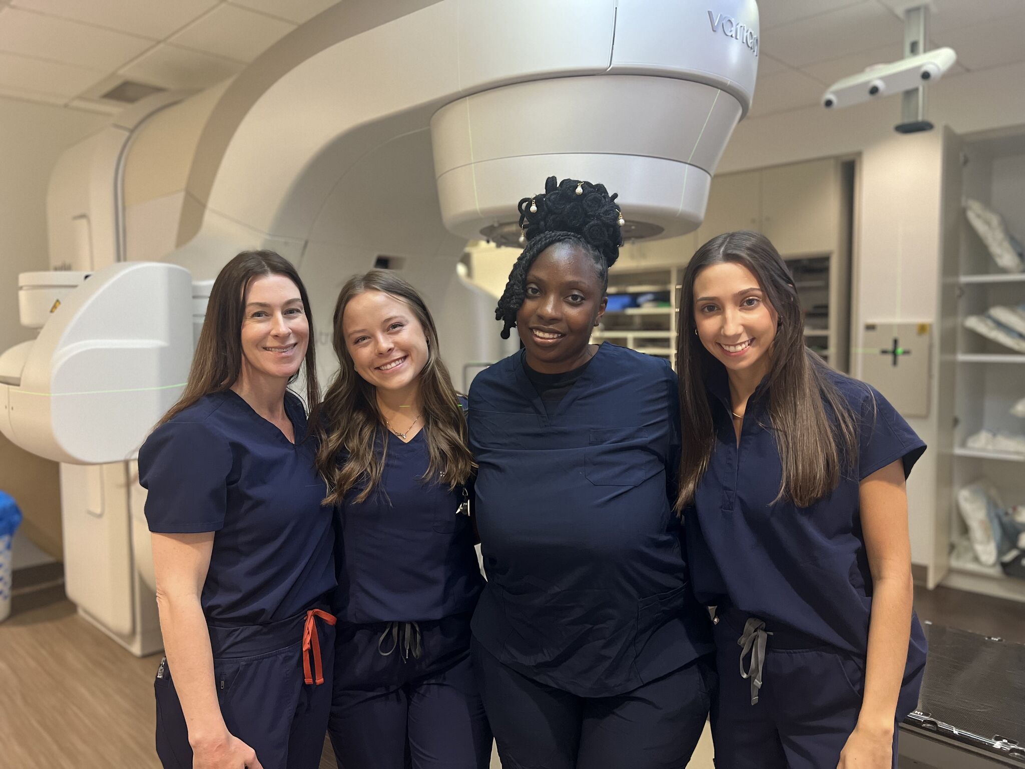
The Role of Diagnostic Radiology in Cancer Care

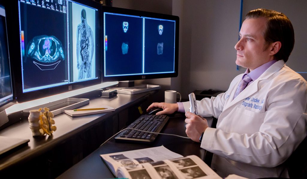
By Dr. Peter Lindley, M.D., Diagnostic Radiologist
From the initial diagnosis that serves as the patient’s entry point into CARTI to continued scans to monitor their response to treatment, diagnostic radiology plays an integral role in the patient’s entire cancer journey. CARTI’s team of diagnostic radiologists utilize the most advanced imaging equipment available – CT, PET, MRI and ultrasound – to gain an in-depth look at what is going on within the patient’s body.
To explain the role of diagnostic radiology, it is important to start at the beginning. Once a medical issue is suspected, the patient’s physician will refer them to CARTI to undergo an imaging study that will identify whether or not cancer is present, and to what degree.
Often, the first test performed is a computed tomography scan, commonly known as a CT. During a CT scan, a special machine uses x-rays to create a series of detailed, computerized pictures of areas inside the body taken from different angles. These three-dimensional images are able to reveal abnormalities in both bone and soft tissues, including organs, muscles and tumors. For most CT exams, a special material called a contrast dye is injected into the patient to make the area of the body being studied easier to see.
The physician may also order a PET/CT scan in order to better delineate overall disease burden or monitor the patient’s response to treatment. During a PET scan, a small amount of radioactive glucose (sugar) is injected into the vein. The PET scanner then takes detailed, computerized pictures of areas inside the body where the glucose is being used. Because cancer cells often use more glucose than normal cells, the pictures can help physicians find cancer cells in the body. These images are then combined with CT scans to create a more complete picture of what is going on in the body than either test can offer alone.
Another common imaging modality is MRI. This can be used as a next step imaging study when questionable findings are discovered on a CT, or it can be used entirely on its own to make an initial diagnosis. MRI uses radio waves and a powerful magnet linked to a computer to create detailed pictures of areas inside the body. These pictures can show the difference between normal and diseased tissue. MRI makes better images of organs and soft tissue than other scanning techniques. But these high-quality images are assured only if the patient is able to remain perfectly still and hold their breath at specific times while the pictures are being recorded. MRI may take more time to perform than other imaging techniques, and some people ― particularly children and people with anxiety or a fear of enclosed spaces ― may find it difficult to lie still throughout the duration of the study. Metal and electronic objects can interfere with the magnetic field of the MRI unit. Patients should tell their technologist if they have medical or electronic devices or other metal objects in their body because they may pose a risk, depending on their nature and the strength of the MRI magnet.
If there is concern for possible bone involvement, CARTI’s imaging team may recommend a bone scan, which is used to examine the bones for damage caused by cancer that either started there or that has spread from another part of the body. For this study, a small amount of radioactive material is injected into the vein then it travels through the bloodstream, collecting in the bones so it can be detected by the scanner.
Ultrasound is often employed to look at soft tissues, especially organs. This safe and painless procedure produces pictures of the inside of the body using high-frequency sound waves. The sound waves are transmitted through a transducer, or probe, that is placed directly on the skin over the area that needs to be examined. The sound waves bounce off internal tissues or organs and form echo patterns that are shown on the screen of the ultrasound machine. Because the images, called sonograms, are captured in real time, they can show the structure and movement of the body’s internal organs, as well as blood flowing through blood vessels.
Once all of the ordered images are received, our diagnostic radiologists closely examine each image and combine those findings with the referring provider’s clinical notes and any relevant lab values and test results in order to create a summary report of the study. This report is then shared with the treating physicians in order to help either diagnose the patient or customize the optimal treatment plan.
To learn more about imaging services available at CARTI, click here. You can also learn more about our team of expert diagnostic radiologists here.