Interventional Radiology Occupying Increasingly Prominent Role in Cancer Patient Care


By Dr. David Hays and Dr. Edgar D. St. Amour

One of the most exciting subspecialties among cancer centers wasn’t originally developed for and isn’t limited to fighting that disease. Interventional radiology (IR), a subspecialty of radiology, uses minimally-invasive image-guided procedures to diagnose and treat diseases in nearly every organ system. It runs a wide gamut of procedures on tumors both malignant and benign. In comparison to open surgery, these procedures have less risk, less pain and less recovery time.
IR was founded in the early 1960s by Dr. Charles Dotter, who invented angioplasty and the catheter-delivered stent, which were first used to treat peripheral arterial disease.
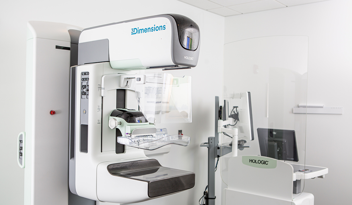
By using advanced imaging in the form of ultrasound, X-rays, CT scans and MRI scans, interventional radiologists can see inside your body and treat complex conditions less invasively and with incredible precision.
Through this subspecialty that is becoming known as the fourth pillar of oncology – along with medical, surgical and radiation – we are occupying an increasingly prominent role in cancer patient care, from initial diagnosis to treatment of malignancy and complications. At the very beginning, when a tumor is detected, we often do the biopsy to determine the diagnosis. A week or two later, if the patient needs chemo or other procedures to treat cancer, we might be involved. We also offer palliative care options – inserting catheters that drain fluids and treating fractures caused by tumors. Basically, we help ease the pain and the burden of the disease itself. Ultimately, IR provides additional treatment options for patients.

One of the procedures in which we might be involved is called ablation. As opposed to surgery, it is a minimally invasive technique that is commonly used in the treatment of tumors of the liver, kidney, bone and lung. It’s a very exciting tool in our toolbox, and in some cases it can be curative. We are able to target tumors directly and either “burn” or “freeze” them. CT, ultrasound or MRI is used to guide and position the needle probe into the tumor.
There is no incision, just a small puncture for the probe. In the liver, for example, we can direct radiation to the precise location. Bed rest is prescribed for a couple of hours, then the patient goes home. All procedures are out-patient, and most patients only require over-the-counter pain reliever. The effectiveness of this new technique in treating cancer depends on the size of the tumor and its accessibility to the probes.
Another technique, called embolization, can be used for tumors that are too big to be treated with ablation.
Radioembolization allows for internal delivery of radiation through the arteries supplying blood to the cancer cells, allowing concentration of high doses of radiation to the cancer with minimal effect on the surrounding healthy tissues. With IR, we can go anywhere in the body, using arteries and veins as a highway for treatment. Depending on the procedure planned, we may go directly into the organ, rather than the blood vessels.
Since its introduction in 2000, interventional radiologists have been the leaders in the use of Y-90 radioembolization to treat liver cancer.
Through this technique, the catheter goes straight to the liver, delivering radiation directly. Although it often requires two treatments, instead of cutting out the portion with cancer, we can take out the segment with radiation. This is a great option for patients who are not good surgical candidates.
When a patient has surgery, they are recuperating for weeks. An IR patient has only two or three days of downtime, and they may go back to other treatment right away. It doesn’t disrupt other therapies as much, and it allows for an integrated continuum of care.
As previously mentioned, not all tumors are malignant. IR can treat uterine fibroids as well. Fibroids can cause pain and bleeding from a woman’s uterus, and traditional treatment has been to remove the uterus. Uterine fibroid embolization is a minimally invasive option that preserves the uterus and greatly reduces the recovery time, compared to surgical procedures. Preserving the uterus means the patient may be able to bear children after the procedure.
Looking forward, we’re excited about how IR will continue to play a role in advances in cancer treatment. Eventually, IR will be involved with immunotherapy, for example.
As doctors at CARTI, we’re all under one roof, and we meet every Thursday for what is essentially a case conference to talk about our patients. We are there every week to offer our IR services as well. It’s a huge benefit to have such a collaborative team – a multidisciplinary team – deliver unparalleled oncology care for the patients of Arkansas.